![]()

![]()
![]()
![]()
![]()
![]()
|
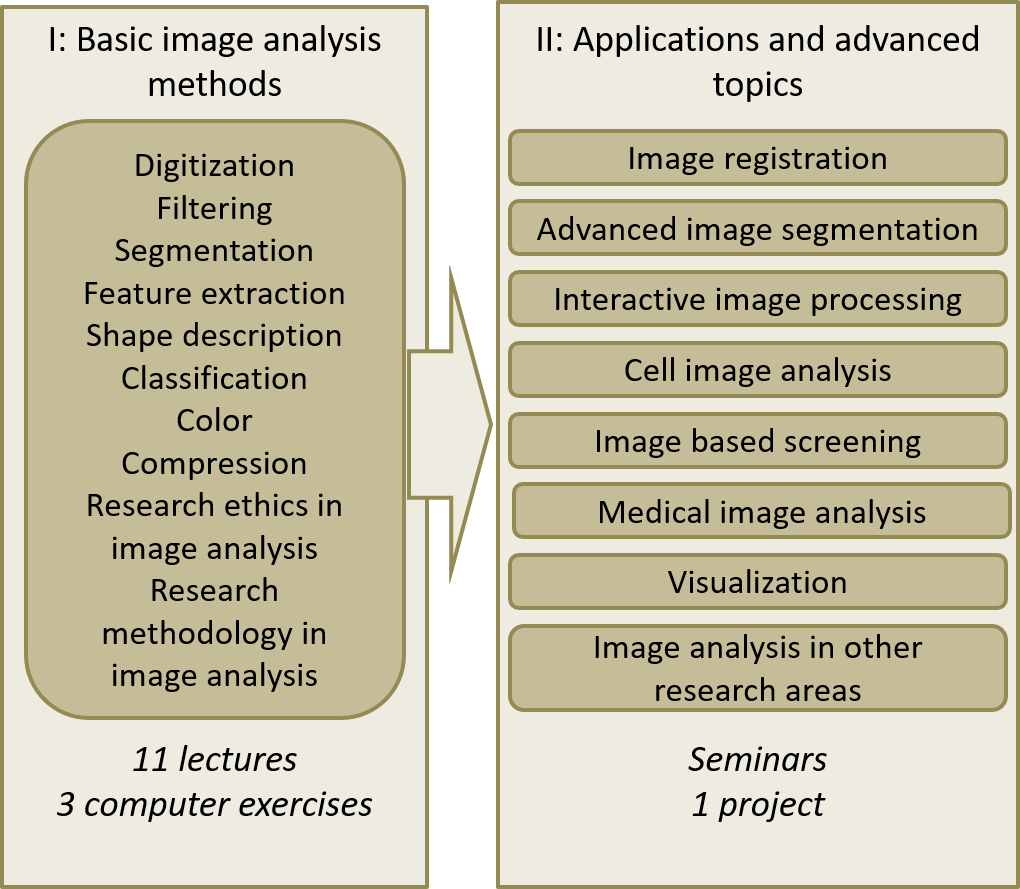
ECTS credits: 8 for the whole course (with a project), 5 for a shorter version Course period: January-February 2022 Lectures and computer exercises will be given online on Tuesdays and Wednesdays (10-16), starting from January 18 to February 23 (weeks 3-8). The course will be given remotely via Zoom and Studium. More information will be sent to registered participants in December. Schedule v1.1 - it may still change! Course aim: This course aims at giving doctoral students and researchers from different disciplines sufficient understanding of digital image processing and analysis techniques to solve basic image analysis problems. The content of the course includes methods especially suitable for MAX IV data. The course will also offer an introduction to several freely available software tools (e.g. CellProfiler, ImageJ and ilastik), preparing the participants to start using computerized image analysis in their research. By inviting researchers interested in using MAX IV and image analysis, and allowing them to work on data of similar type but produced elsewhere, the course has the added value of attracting more researchers to MAX IV and also increasing the awareness of future possibilities giving a head-start in defining and planning MAX IV projects. The basics of image analysis are general, but by designing examples and hands-on computer exercises based on published data from other world-leading X-ray facilities, we can prepare participants for the future. After the course, the participants will not only have a better understanding of the underlying theory and possibilities of image analysis but also be better at designing their experiments in the MAX IV environment. Contents and study format: The focus of the course is on reaching a broad understanding of digital image analysis and a basic understanding of the theory and algorithms behind the methods. The course starts with basic image analysis methods and hands-on computer exercises, including research methodology and related research ethics. The three hands-on computer exercises are designed to get familiar with the interfaces of common software and to solve realistic image processing problems. In the second (optional) part of the course, the course participants will be divided into small groups that focus on a project with one specific data type or application. The course will end with the course participants briefly presenting what they did with the data in their projects, and brainstorming on potential next steps and future possibilities. As part of the course, we are planning guest lectures by Anders Bjorholm Dahl, Professor at the Technical University of Denmark (DTU) and head of QIM: The Center for Quantification of Imaging Data from MAX IV, Oxana Klementieva, Associate senior lecturer at Medical Microspectroscopy - Lund University, and Alexandra Pacureanu, a research scientist responsible of nanoscale X-ray neuroimaging at ESRF, the European Synchrotron. The examination will be divided into:
Target group/s and recommended background: The target group is graduate students and senior researchers from all subjects where computerized image analysis is (or could be) used as a research tool, in combination with MAX IV data. The limit of participants is 20. Application from course participants should be sent to Damian Matuszewski, damian.matuszewski@it.uu.se, not later than December 15. Course coordinator: Damian Matuszewski, damian.matuszewski@it.uu.se Centre for Image Analysis, Division of Visual Information and Interaction Dept. of Information Technology, Uppsala University. Detailed content for the 5 credits course Detailed content for the 8 credits course Detailed course information |
 |
The course is organized in a collaboration between the Division of Visual Information and Interaction at Uppsala University (Vi2), MAX IV, BioImageInformatics facility at SciLifeLab, and National Microscopy Infrastructure (NMI).